
Root Canal Treatment
Microscopic Root Canal Treatment
Comprehensive information about Microscopic Root Canal, treatment steps and FAQs — Dentsmile.
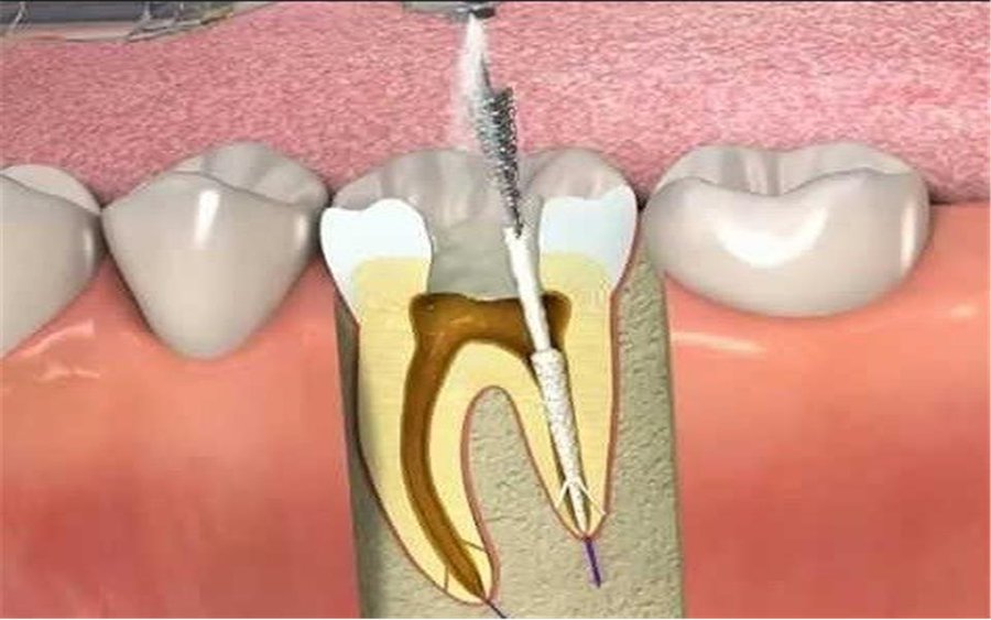
Microscopic root canal treatment is an endodontic treatment approach in which root canal therapy is performed using a dental operating microscope that provides magnification and powerful illumination. The microscope can offer important advantages particularly in cases involving complex root canal anatomy, difficult-to-locate canal openings, or previously treated teeth requiring root canal retreatment.
Root canals are extremely small structures and can be anatomically complex in certain teeth. In addition to the main root canals, a tooth may contain accessory canals, narrowed or calcified canals, and anatomical variations that can be difficult to identify with the naked eye.
A dental microscope magnifies and illuminates the treatment area, helping the dentist evaluate these structures in greater detail.
However, the use of a microscope alone does not guarantee successful root canal treatment. Accurate diagnosis, knowledge of root canal anatomy, appropriate disinfection and shaping techniques, proper root canal filling, and an adequate final restoration all contribute to the overall success of treatment.
When Is Microscopic Root Canal Treatment Used?
A microscope can also be helpful during routine root canal treatment. However, the magnification and illumination it provides become particularly valuable in difficult and complex endodontic cases.
Microscopic root canal treatment may be particularly useful in the following situations:
Difficult-to-Locate Root Canals: Some teeth may contain additional or very narrow canal openings. A microscope can help these areas be examined in greater detail.
Calcified Canals: Root canals may become narrowed due to age, trauma, or other factors, making them more difficult to locate in certain areas.
Complex Root Canal Anatomy: The number and shape of roots and canals vary between individuals. Magnification can help evaluate these anatomical details.
Root Canal Retreatment: In teeth that have previously undergone root canal treatment but continue to show signs of infection, magnification can provide important advantages when evaluating existing materials and locating canal entrances.
Separated Instruments Within the Canal: If an endodontic instrument has fractured inside a root canal, its position and the possibility of removing it can be assessed more precisely under magnification.
Assessment of Perforations: Magnification and illumination may assist in identifying and managing certain perforations involving the root or pulp chamber floor.
Teeth with Suspected Cracks: A microscope can help provide a more detailed assessment of certain cracks on the tooth surface. However, it should not be assumed that every crack can be definitively identified under a microscope.
Apical Surgery: Dental microscopes may also be used during endodontic surgical procedures such as apical resection to provide a more detailed view of the root tip and surrounding structures.
How Is Root Canal Treatment Performed Under a Microscope?
The fundamental stages of microscopic root canal treatment are similar to those of conventional root canal therapy. The main difference is the use of high magnification and illumination during some or all stages of the procedure.
Examination and Diagnosis
The patient’s symptoms are first discussed, and the affected tooth is clinically evaluated.
Radiographic images are reviewed, and pulp vitality tests may be performed when necessary. In complex cases, three-dimensional imaging methods such as cone beam computed tomography (CBCT) may also be considered.
The microscope and CBCT are not alternatives to one another. The microscope provides direct magnified visualization of the treatment area, while CBCT can provide three-dimensional information about the tooth and surrounding structures when clinically indicated.
Local Anesthesia
Local anesthesia is administered to the treatment area to help keep the patient comfortable during the procedure.
Tooth Isolation
Isolating the tooth from the oral environment is important during root canal treatment. When appropriate, a rubber dam is used to separate the treated tooth from saliva and microorganisms within the mouth.
Locating the Root Canal Openings Under Magnification
After access to the pulp chamber has been established, the canal openings are evaluated using the magnification and illumination provided by the microscope.
Searching for additional canals that might otherwise be difficult to identify is particularly important in teeth with complex anatomy.
Cleaning and Shaping the Root Canals
Infected or damaged tissue is removed from the root canals. The canals are cleaned, disinfected, and shaped using endodontic instruments and appropriate irrigation solutions.
A microscope does not provide a direct view of the entire length of every root canal. Deep portions of curved canals, in particular, may not be directly visible. For this reason, radiographic assessment, electronic apex locators, and other endodontic methods continue to play an important role during treatment.
Filling the Root Canals
Once the canals have been cleaned and shaped, they are filled using appropriate root canal filling materials.
The aim is to seal the root canal system as effectively as possible and reduce the risk of microorganisms re-entering the canals.
Restoring the Tooth
After root canal treatment, an appropriate final restoration such as a filling, onlay, or crown may be planned according to the amount and condition of the remaining tooth structure.
The long-term outcome of root canal treatment depends not only on the procedures performed inside the root canals but also on restoring the tooth appropriately afterward.
What Are the Advantages of Microscopic Root Canal Treatment?
A dental operating microscope can provide important visual advantages, particularly in complex cases.
The main advantages include:
High Magnification: Helps provide a more detailed view of very small anatomical structures.
Powerful Illumination: Improves visualization of the pulp chamber and root canal openings.
Assistance in Locating Additional Canals: Can make it easier to identify additional canal entrances, particularly in certain molar teeth.
More Controlled Treatment: May help reduce unnecessary removal of healthy tooth structure.
Advantages During Root Canal Retreatment: Can facilitate assessment of previous restorations, root canal filling materials, and potential complications.
Assistance with Separated Instruments and Perforations: In suitable cases, magnification can contribute to more controlled management of these complications.
Is Microscopic Root Canal Treatment More Successful?
A dental microscope is an important tool that allows the dentist to see the treatment area under greater magnification and improved illumination. Particularly in complex cases, it may contribute to more controlled treatment.
However, it is not accurate to assume that “if a microscope is used, the root canal treatment will definitely be successful.”
The success of root canal treatment depends on factors including:
Accurate diagnosis
Root canal anatomy
Extent and nature of the infection
Cleaning and disinfection of the canals
Quality of the root canal filling
Amount and condition of remaining healthy tooth structure
Quality of the final restoration
Patient’s overall oral and dental health
The microscope is one of several technologies that can help make this process more precise and controlled.
What Is the Difference Between Microscopic and Conventional Root Canal Treatment?
The fundamental objective is the same in both approaches: to remove infected or irreversibly damaged pulp tissue, clean and disinfect the root canal system, and fill it appropriately.
The main difference in microscopic root canal treatment is that the dentist can work under high magnification and powerful illumination.
This can become particularly important when dealing with anatomical details that are difficult to identify under conventional visualization.
Microscopic root canal treatment is therefore not an entirely different type of root canal therapy. It is root canal treatment performed with the additional visual control provided by a dental operating microscope.
Can a Previous Root Canal Treatment Be Redone Under a Microscope?
Yes. One of the important applications of dental microscopy is root canal retreatment.
A previously root canal-treated tooth may present with:
An untreated additional canal
Persistent infection
Existing materials within the canals
A separated endodontic instrument
Perforation
Complex root canal anatomy
A microscope can provide significant advantages when evaluating and managing these situations.
However, not every unsuccessful root canal treatment can or should be retreated. The tooth must also be considered maintainable from restorative and periodontal perspectives.
Can a Separated Root Canal Instrument Be Removed Under a Microscope?
The fine endodontic instruments used during root canal treatment can occasionally fracture within the canal.
A dental microscope can help determine the location of the separated instrument and, in suitable cases, assist with its removal.
However, not every separated instrument can or needs to be removed.
The location of the instrument, root anatomy, remaining tooth structure, and the amount of root tissue that would need to be removed to retrieve the instrument should all be considered.
In certain cases, attempting to remove the separated instrument may cause more damage to the tooth than leaving it in place. The treatment decision should therefore be based on an individualized risk-benefit assessment.
How Many Appointments Does Microscopic Root Canal Treatment Require?
The number of appointments depends more on the condition of the tooth and the infection than on whether a microscope is used.
Some cases can be completed in a single appointment, while complex anatomy, retreatment cases, or certain types of infection may require multiple visits.
It is therefore not appropriate to provide an exact treatment duration without clinical and radiographic assessment.
Is Microscopic Root Canal Treatment Painful?
As with conventional root canal treatment, microscopic root canal therapy is generally performed under local anesthesia.
When adequate anesthesia has been achieved, significant pain during treatment is not usually expected.
Mild sensitivity or discomfort when biting may occur for several days after treatment. Sensitivity may be more noticeable when infection was already present around the root before treatment.
A dentist should be contacted if pain becomes increasingly severe, significant swelling develops, or unexpected symptoms occur.
Microscopic Root Canal Treatment in Üsküdar, Istanbul
At Dentsmile Oral and Dental Health Clinic – Üsküdar, Istanbul, when evaluating a patient for microscopic root canal treatment, we first consider the current condition of the tooth and whether the natural tooth can reasonably be preserved.
Working under magnification can provide important advantages particularly in teeth with complex root canal anatomy, previously treated teeth with persistent symptoms, difficult-to-locate canals, and certain endodontic complications.
However, the use of a microscope is not, by itself, an indication for treatment. Every patient should first undergo clinical and radiographic evaluation, with three-dimensional imaging considered when necessary.
When planning microscopic root canal treatment in Üsküdar, the fundamental objective is not simply to use advanced technology, but to combine accurate diagnosis with appropriate endodontic techniques to preserve the natural tooth whenever reasonably possible.
More intervention does not necessarily mean a better outcome in microscopic endodontics. Particularly when working with narrow or calcified canals, it is important to progress carefully without unnecessarily removing healthy tooth structure.
At Dentsmile, our approach to microscopic endodontics is to benefit from the advantages of magnification and illumination while preserving as much healthy tooth structure as possible and treating the tooth with the aim of maintaining long-term function.